The first milestone of a bionic artificial neural network.
Editor’s note: This article is from WeChat public account “ Quabit ” (ID: QbitAI ), Author thirteen, Bian Ce, Yu Yang.
Brain exploration, go one step further today.
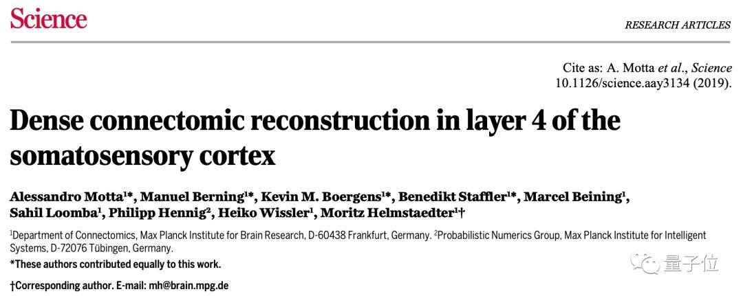
The latest Science magazine cover has published the latest brain science achievements of the well-known German Max Planck Brain Institute:
They sharpened a sword in seven years, reconstructed a very complex cerebral cortex neural network, and revealed the largest mammalian neural circuit diagram to date.
Before, humans only knew the “look” of brain neurons. Now, how mammalian neurons are connected-for the first time has been revealed, and a larger number of cerebral cortex neural networks have been reconstructed.
Also, the method of AI plays an important role in it. The researchers also said that this breakthrough may further provide guidance for the development of AI:
Uncovering the secrets of biological neural network connection may further explore the principle of efficient computing in the brain. This is the first milestone in the trend of artificial neural networks that are constantly learning from biological neural networks.
So what kind of groundbreaking research is this?
Unveiled neuronal connections in mammalian brains for the first time
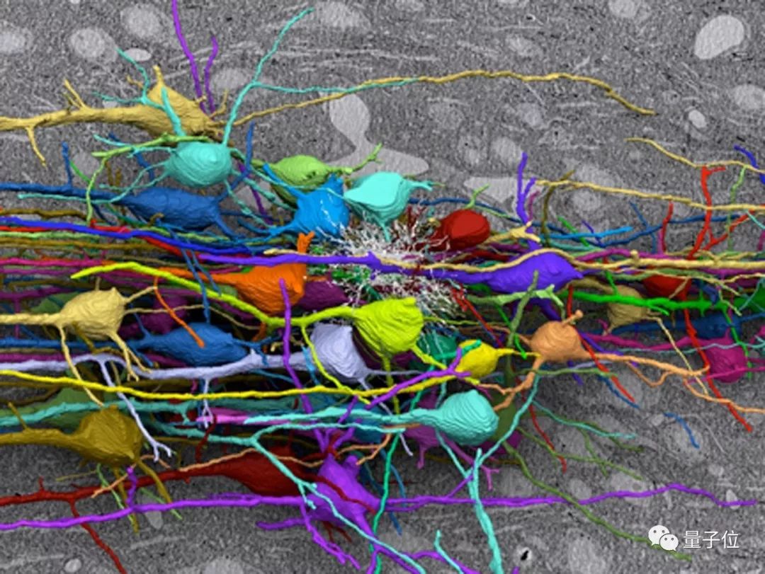
The mammalian cerebral cortex is a very complex network of neural processes: long, thin, branched, and very dense.
This high packing density makes the reconstruction of cortical neural networks quite challenging.
Previous studies have focused on overall imaging, but this time, scientists’ reconstruction work has really penetrated into the connection of neurons.
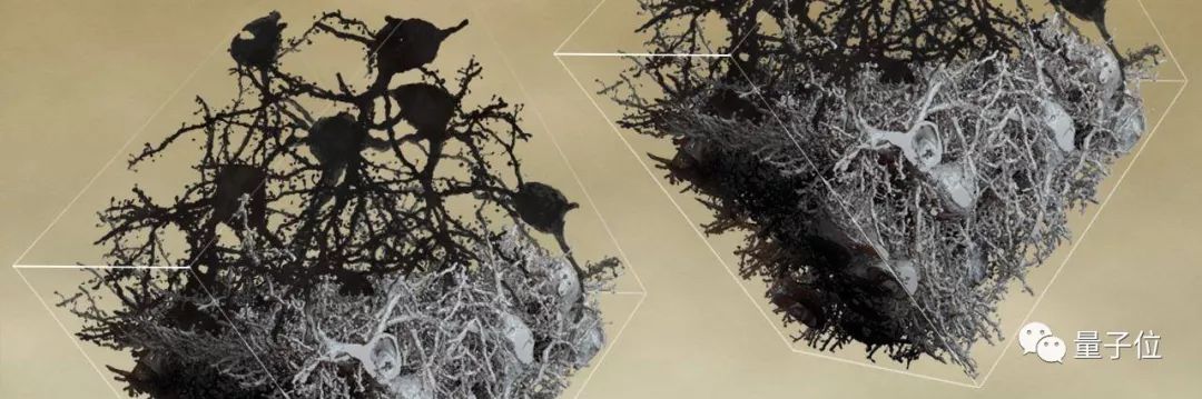
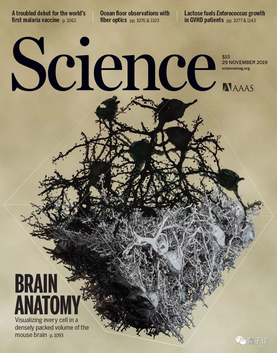
Researchers from the Max Planck Institute for Brain Research in Germany used artificial intelligence to reconstruct the morphological characteristics and connections of 89 neurons in the mouse barrel cortex with high spatial resolution.
Also, the area covered by this study is two orders of magnitude larger than the methods attempted by early neuroanatomical mapping, and is 300 times the volume of the dense reconstruction of the mammalian cerebral cortex.
Corresponding author Moritz Helmastaedter), the findings of this study reveal the largest group of mammalian neural connections to date.
In addition, through the analysis of the connection group circuit, this kind of research breakthrough in biological intelligence is likely to migrate to the AI field, which will have a significant impact on artificial intelligence.
Moritz said:
Mapping the neural network in the cerebral cortex is a major scientific adventure. We want to uncover the truth about the efficient operation of the brain as a computing machine. Its model is so different from today’s AI.
Besides, there are some amazing details: the connection group data can extract inhibitory and excitatory neuron subtypes whose geometric information cannot be predicted.
The research team believes that applying their method to cortical tissues of different brain regions, cortex, development time points, and species can reveal how natural evolution designed the biological warp network and neural network. How fine-grained structures are shaped.
In addition, screening of the connected group can reveal the circuit phenotype of neuropathy and related brain diseases, telling us how important certain brain diseases are affected by the connected group and neural circuits.
Reconstruction of the strongest cerebral cortex neural network
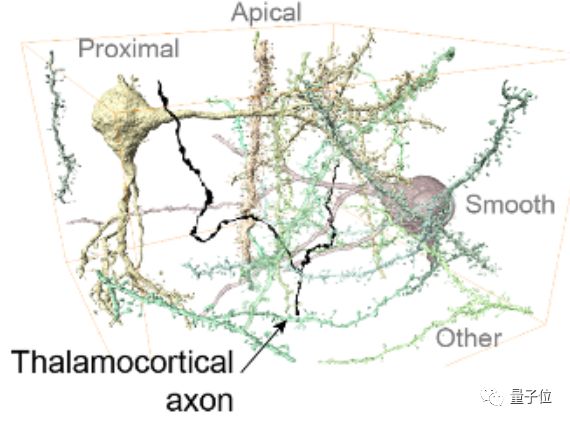
The mammal’s brain consists of a very dense network of neurons, including axons and dendrites of nerve cells.
The density of these nerve cells is very high. In the past, optical imaging methods could only distinguish a small part of the nerve cells in the mammalian cerebral cortex.
The development of three-dimensional electron microscope technology has made it possible for researchers to draw stereoscopic images of neuron structures.
Although the program speed of this microscopy technique has been greatly improved, reconstruction of 3D images from 2D images has been prone to errors in the past, resulting in limited analysis of 3D image data.
Now, AI-based methodsPlayed an important role.
Researchers integrate human data analysis into neural connection data generation, and use the efficiency of human-machine data analysis to promote the progress of neural connection group.
The way they improve efficiency is as follows:
-
1. Improve the quality of automatic segmentation;
-
2. Analyze the possible wrong positions in automatic segmentation and guide manual work to these positions;
-
3. By helping the annotator to optimize personnel data interaction, realize the fast transmission of internal parallel data, and minimize the delay between the annotation program queries.
After optimization, nearly 100 student annotators can solve thousands of reconstruction problems in 29 seconds.
In the end, it took them about 4,000 working hours to reconstruct a total of 2.7 meters of neuronal filaments in the fourth layer of the mouse somatosensory cortex.
The cortical data reconstructed by this work is 300 times larger than before, and the efficiency is increased by 20 times.
They analyzed the junctions between 6,979 presynaptic membranes and 3,719 postsynaptic membranes, each synapse being connected to at least 10 synapses, for a total of 153,171 synaptic connections, and then analyzed the Dense circuit structure.
Using human-computer interaction to analyze the connected group of neuronal tissue, researchers have obtained the largest connected group data of the cerebral cortex to date.
Using these data, the researchers established a connection phenotype analysis method for the local dense neuron circuit of mammalian cerebral cortex, which provided the possibility to screen from the connection group between various neural tissues.
So how exactly was the experiment designed and carried out?
Interpretation of experimental methods
Specifically, the researchers first sampled and stained mouse tissue.
48 hours after fixation, the brain was removed from the skull and its coronal sections were cut using a fiberglass knife.
Using a 1-mm biopsy puncture machine, two samples were taken from a slice 1 mm thick 5 mm from the front of the brain. The target was the fourth layer of the somatosen-sory cortex in the right hemisphere.
The extracted tissue was then stained and hardened at 60 ° C for 48 hours.
This is followed by a 3D electron microscope experiment.
Place the embedded sample on an aluminum stub and trim it so that the tissue can be directly exposed on all sides of the sample.
The sides of the sample were coated with gold using a sputter coater and placed in a SBEM device.
△ SBEM: A continuous block scanning electron microscope is a method for generating high-resolution three-dimensional images from small samples.
In the EM overview image, the transition between L4 and L5A is identified by a sudden drop in somatic cell density between the two layers (Figure 1C).
During this process, a total of 3420 image planes were collected, for a total of 194GB of data.
The next step is image alignment.
After acquiring the 3D EM data set, manually inspect all images and mark imaging artifacts caused by fragments appearing on the sample surface during the imaging process.
Images with fragmentation artifacts have been replaced on images with the same location from the previous or next plane.
The following modifications are applied:
When obtaining a shift vector with an offset of more than 100 pixels, manually reducing the weight of the corresponding entry in the least squares relaxation by 1000 times (until the highest residual error is less than 10 pixels) To iteratively correct these errors.
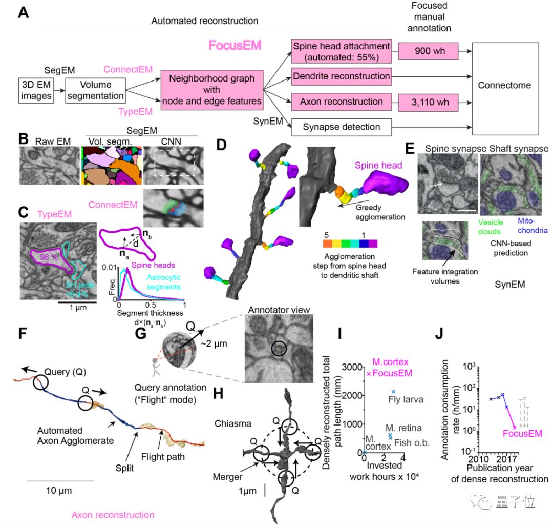
The following figure is an effective way to reconstruct the dense connection group (connectomic).
△ A method for effectively reconstructing dense connected groups.
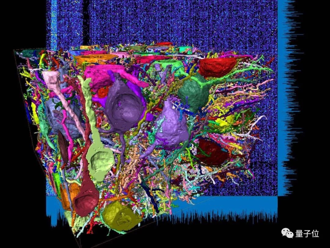
FirstUsing an automated heuristic to detect blood vessels and cell bodies, and then using machine learning-based image segmentation to process the remaining image volume, the result of this processing is 15 million fragments corresponding to axons, dendrites, and somatic cells.
Based on this, a neighborhood graph between segments is constructed, and the interface properties between directly adjacent segments are calculated.
Based on these characteristics, the researchers trained a connection classifier (ConnectEM, A and B in the figure above) to determine whether two fragments should be connected, or whether they should be disconnected.
Using the SynEM classifier, the researchers determined whether an interface between the two separation processes corresponds to a chemical synapse, and if so, which one is a pre-synapse and which is a post-synapse.
The next step is the reconstruction of cellular neurons.
Researchers used a simple set of growth rules to automatically connect neurites, which are based on a fragment-to-fragment proximity map and a connection and neurite type classifier.
The result is a fully automatic reconstruction of the somatic and dendritic processes of neurons.
For 89 cells, with only 9.7 hours of additional manual correction, the dendritic axes of these new molecules can be reconstructed without merging errors, while 37 division errors are retained, and the dendritic length recall rate is 87.3 %.
This is followed by dense tissue reconstruction.
Rebuilding neurons from the cell body is not a major challenge.
Axons and dendrites are not connected to the cell bodies in the data set, and are densely distributed in the tissue, accounting for about 97% of the total neuron path length in this part of the cortex (G in the figure above).
To reconstruct the vast majority of neurites (H in the image above), the researchers used their connectivity and neurite type classifiers (ConnectEM and TypeEM) to merge neurite fragments into larger dendrites And axonal agglomerate.
Researchers also reconstructed synaptic detection, post-synaptic target types, and linkers.
Considering pre-synaptic and post-synaptic neurons reconstructed in tissue, the researchers also extracted their connectors. To this end, SynEM is used to detect synapses between axonal and presynaptic synapses.
It is worth mentioning that the team also trained a specialized axonal interface classifier using training data containing only axonal and soma synapses.
There is no doubt that the great progress made by so many methods is also the result of cross-disciplinary and cross-innovation.
Interdisciplinary research team
This research work lasted for 7 years, and the research team came from the Max Planck Brain Institute in Germany.
The four common works are:
Alessandro Motta is a PhD student at the Max Planck Institute for Brain Research, and is a student of the corresponding author Moritz Helmastaedter.
Manuel Berning is a physicist, neuroscientist, and a programmer. He studied at the Max Planck Brain Institute from 2014 to 2017 and is currently a data scientist at SAP (German Software Corporation).